Shoulder external rotation at side
Medial Tibial Stress Syndrome (MTSS) or “shin splints” is one of the most commonly reported lower limb injuries by competitive and recreational athletes. Recent research has shown that shin splints affects approximately 20% of the running population, with the majority of sufferer’s partaking in long distance training/competition.
Currently, there is two widely accepted theories on the cause of shin splints:
The bony bending theory suggests that during running, the Tibia (shin bone) bends due to the stress placed upon it. This bending causes small amounts of strain in the bone that enables it to adapt and get stronger (a good thing!!). When this strain exceeds the adaption process the shin bone becomes overloaded (a bad thing!), subsequently leading to injury and pain.
The traction theory states that shins splints is caused by the continual contraction of the muscles (Soleus, Flexor Digitorum Longus & Tibialis Posterior) that attach to the inner border of the shin. As these muscles contract during running, they place a traction stress on the shin bone, which results in inflammation at their attachment onto the bone, causing pain.
Current research has identified several risk factors leading to an increased likelihood of developing shin splints. These include:
A previous history of shin splints
High Body Mass Index (BMI)
Female gender
Decreased running experience
Decreased running cadence (step rate)
Excessive pronation
Crossover running style
Increased vertical oscillation (ground clearance)
Forefoot running
To diagnose shin splints accurately, two symptoms must be present:
If you are experiencing symptoms not typical of shin splints such as cramping, pain spanning less than 5cm, burning pain, numbness or pins and needles, you should seek a thorough assessment by a physiotherapist to properly diagnose and treat your condition.
Arguably one of the biggest contributors to the development of shin splints in a runner is their running technique, particularly their lower limb mechanics. One of the quickest ways to reduce shin splints related pain is to address the technical aspects of running that can contribute to increased stress across the Tibia and associated musculature. What you should focus on is:
Increase your cadence!! – This is by far the biggest bang for your buck. Increasing your cadence by approximately 10%:
Reduces lower limb impact forces by 20%
Reduces ground contact time
Reduces stride length
The best way to achieve an increase in your cadence is by using GPS watches, phone applications or by simply running on a treadmill.
Eliminate a crossover running style – On a track, run straddling a line across 2 lanes or alternatively, try and maintain a space between your knees with every stride.
Strength exercises for shin splints should aim to improve the localised muscular capacity of the calf complex as well as the bone load capacity of the Tibia. This is best addressed with weight bearing functional exercises that mimic running postures.
One of the most important and often forgotten muscles of the calf complex is the Soleus. The soleus muscle is vital for absorbing excessive loads placed on the Tibia during running by minimising excessive pronation as well as resisting the bending forces experienced by the Tibia due to ground impact.
The best Soleus exercise that runners can do is the Bent Knee calf raise (pictured above). To perform the exercise correctly:
Bend your knee as far forward as possible, keeping your foot flat on the floor
Lower your heel back to the ground
Perform 3 sets of 15 repetitions in a slow and controlled manner.
As always, if you are having problems, please do not hesitate to contact one of our experienced physiotherapists.
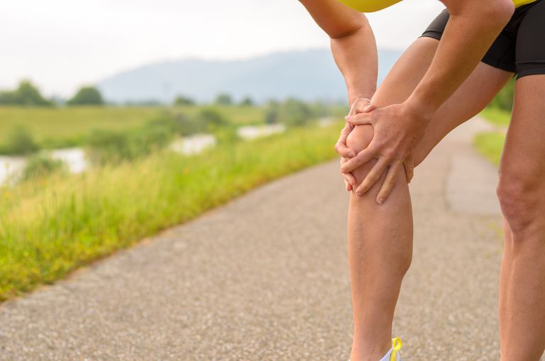
Patellofemoral Pain Syndrome (PFPS) or “Runners Knee” is one of the most common overuse injuries amongst the active population. PFPS accounts for approximately 15% of all knee pain, with females and young adults being 2 times more likely to develop symptoms due to PFPS (Boling et al, 2010).
The Patello-femoral joint is one of two joints that make up the knee (see figure 1). It is comprised of the kneecap (patella) and the thigh bone (femur) and provides the attachment sites for our quadriceps and patella tendons.
PFPS is characterised as “pain experienced around or behind the knee cap, which is aggravated by weight bearing activities that require a flexed knee such as squatting, running, jumping and hopping” (Crossley et al, 2016). It is not uncommon to also experience symptoms such as:
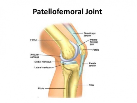
Figure 1. Patello-Femoral Joint
Runners often develop PFPS due to a combination of several factors such as:
Muscular weakness (Quadriceps/Glutes)
Muscular tightness
Changes to training loads
Inappropriate footwear
Anatomical variations in knee cap shape/position
Changes to running style
Biomechanical abnormalities
Currently, the best treatment supported by research is STRENGTH!!!
Strength interventions have been shown to be most effective in relieving pain and improving function in individuals with PFPS. Exercise selection should be patient specific and target the hip external rotators/hip abductors (Glut Medius) as well as the knee extensor muscles (Quadriceps) due to their roles in knee biomechanics (Martin et al, 2018).
The great news is approximately 90% of individuals suffering from PFPS will be completely symptom free within 6 weeks of starting a strength rehabilitation program guided by a physiotherapist.
Amongst the literature there is a lack of strong evidence, supporting the long-term use of:
Electrical Stimulation
Ultrasound
TENS (Transcutaneous Electrical Nerve Stimulation)
Massage
Biofeedback devices
Taping
Orthotics
Dry needling
Acupuncture
Although the majority of these things can help your pain in the short term, none have been shown to be superior to strength exercises of the quadriceps and gluteal musculature.
Research on running mechanics has shown that stress on the Patello-femoral joint is greatest during mid stance and exceeds approximately 7x your body weight. This can be largely increased with
Slower speeds of running (Increase knee flexion)
Large vertical oscillation
Cross over style running
Excessive body twisting
Excessive body twisting
Ways in which you can alter your running technique to reduce knee joint stress by 15-20% are:
Maintain space between your knees when running (knee window)
Increase running cadence by 10%
Minimise excessive body twisting
Currently the best evidence for preventing future patella issues besides maintaining your strength is by assessing and monitoring acute: chronic training volumes in term of distance covered (Km). Acute increases in training volume should be no greater than a factor of 1.5 or you place yourself at an increased risk of suffering a subsequent injury.
We have covered this in depth in our blog titled Our Top 3 injury prevention strategies, which can be accessed by the link below:
Top 3 Injury Prevention Strategies + Lessons from Leicester City
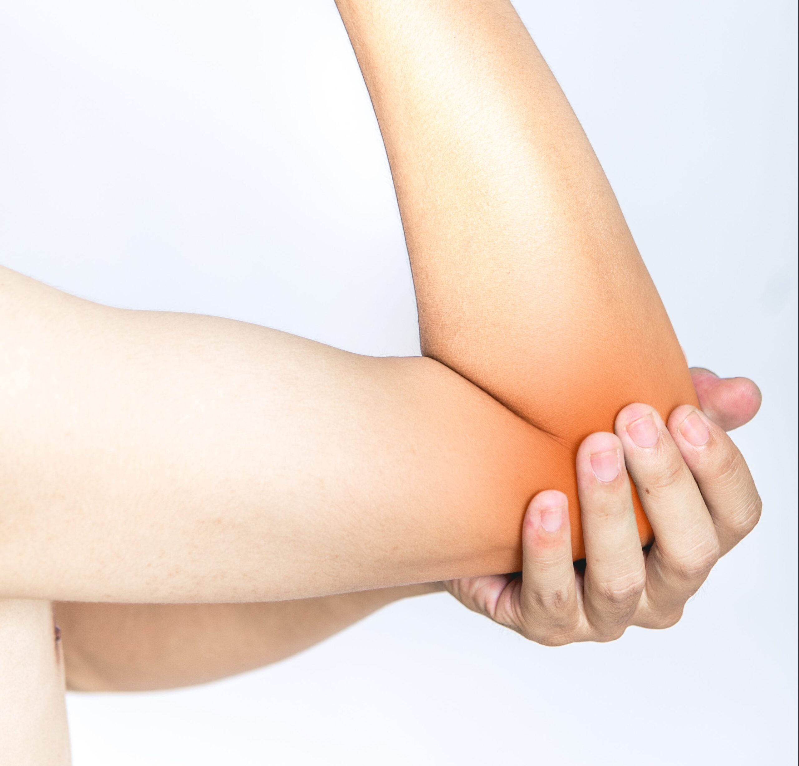
It is an overuse injury from repetitive or forceful/explosive movements involving eccentric motion and/or in which the wrist frequently deviates from a neutral position. This can be from training errors, inadequate equipment or poor environmental conditions.
Tennis elbow can affect anyone, however is more common in people between 30 and 60 years of age. It appears to be more severe and of longer duration in females. The most commonly affected arm is the dominant arm. It is commonly seen in office workers (repetitive typing) or manual labour workers (carpenters etc).
Pain and tenderness over the elbow bone (lateral epicondyle)
Pain with gripping, twisting, lifting.
Some cases may have nerve involvement – nerve pain and neck range of motion restrictions.
A diagnosis can be made based on the history of the condition and a physical examination. X-rays may be used to help rule out other causes of elbow pain, such as arthritis. An ultrasound or magnetic resonance imaging (MRI) scan will show the degenerative changes or small tears in the tendon, but is rarely required.
Evidence tells us that strength exercises are the most effective way of treating tennis elbow, with adjuncts of manual therapy (lateral elbow glides and C5 glides if radial nerve involvement. (L.Bisset et al 2015, Cleland et al 2013).
Strength exercises can not only help settle the pain, but also reduce the risk of the pain returning.
CLICK HERE TO VIEW THE 2 EASIEST, AT HOME STRENGTH EXERCISES!
Each patient should be treated based on the history and the findings. Common treatments include:
Patients can also be reassured that some cases will improve without intervention and just information regarding modification of aggravating activities, ergonomic advice and reassurance that their condition will eventually settle.
Corticosteroid injections are NOT recommended. In a study by Vicenzino et al 2006, 198 participants got assigned to three groups (physiotherapy interventions, corticosteroid injections and ‘the wait and see approach’). The corticosteroid group had most reported recurrences at 72%.
Can provide short term pain relief, however has no effect on long term outcome.
There is conflicting evidence for the effectiveness of bracing/taping compared with placebo or no treatment.
Conflicting evidence, but may be more effective than placebo and ultrasound at relieving pain and improving self-assessed treatment benefit in the short term.
May be beneficial in short term compared with placebo, likely no difference between laser and other active interventions in the short or long term.
No more effective than placebo for pain relief or self-perceived global improvement in short term.
Little or no benefit in reducing pain or improving function.
No benefit.
If you have any questions, or would like our help, please do not hesitate to get in touch at clinicalphysiostives.com.au
1) Physiotherapy management of lateral epicondylalgia – Bisset, Vicenzino (2015)
•Hypoalgesic and sympathoexcitatory effects of mobilization with movement for lateral epicondylalgia – Paungmali, O’Leary, Souvlis, Vicenzino (2003)
2) Specific manipulative therapy treatment for chronic lateral epicondylalgia produces uniquely characteristic hypoalgesia – Vicenzino, Paungmali, Buratowski, Wright (2001)
•Manipulation of the wrist for management of lateral epicondylitis: A randomized pilot study – Struijs, Damen, Bakker, Blankevoort, Assendelft, Van Dijk (2003)
3) Incorporation of Manual Therapy Directed at the Cervicothoracic Spine in Patients with Lateral Epicondylalgia: A Pilot Clinical Trial – Cleland, Flynn, Palmer (2013)
4) A randomized controlled trial of eccentric vs. concentric graded exercise in chronic tennis elbow (lateral elbow tendinopathy) – Peterson, Butler, Eriksson, Svardsudd (2014)
5) Mobilisation with movement and exercise, corticosteroid injection, or wait and see for tennis elbow: randomised trial – Bisset, Beller, Jull, Brooks, Darnell, Vicenzino (2006)
6) Addition of isolated wrist extensor eccentric exercise to standard treatment for chronic lateral epicondylosis: A prospective randomized trial – Tyler, Thomas, Nicholas, Malachy, McHugh (2010)
7) Is tendon pathology a continuum? A pathology model to explain the clinical presentation of load-induced tendinopathy – Cook & Purdam (2009)
Advanced rotator cuff exercise to improve strength of the external rotators (supraspinatus, infraspinatus and teres minor).